


*Culture of adherent cells on a 3D gel matrix under flow conditions
*Transendothelial migration studies under flow conditions
*Establishment and microscopic read-out of endothelial barrier assays (without any artificial membrane)
*Simulation of blood vessels with endothelial cell culture on a soft substrate
*Long-term or short-term cell culture perfusion experiments with defined shear stress and optimal nutrient supply into 3D structures
*Apical-basal cell polarity assays
*Cell barrier model assays with apical-basal gradients
*Cell boundary assays without filters or porous membranes
*Rolling and adhesion assays using leukocytes
*Inflammation studies
*Transport studies
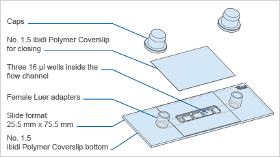
specifications:
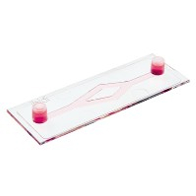
Outer dimensions (w x l) | 25.5 x 75.5 mm2 |
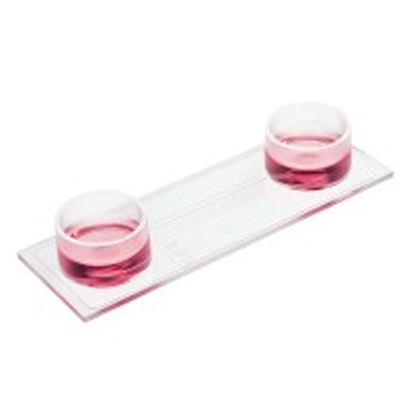
Number of wells | 3 |
Volume of wells | 16 μl |
Well dimensions | 5.4 mm x 4.0 mm |
Well height (without channel) | 0.8 mm |
Growth area per well | 0.21 cm2 |
Coating area per well | 0.34 cm2 |
Channel width | 5.0 mm |
Channel volume (without wells) | 150 μl |
Channel height (without well) | 0.6 mm |
Adapters | Female Luer |
Volume per reservoir | 60 μl |
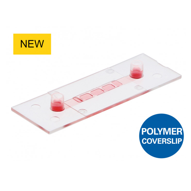
Top cover: | |
Bottom: |
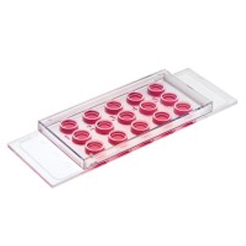
* Can create low or high shear stress in the 0.6 mm channel
* Initial open well format allows for easy sample preparation, then is simply closed with a coverslip for the application of flow
* The well design corrects meniscus formation and allows for superb cell visualization in one focal plane
* Can be used with all ECM hydrogels
(e.g., Collagen Type I, Matrigel, Fibrinogen)
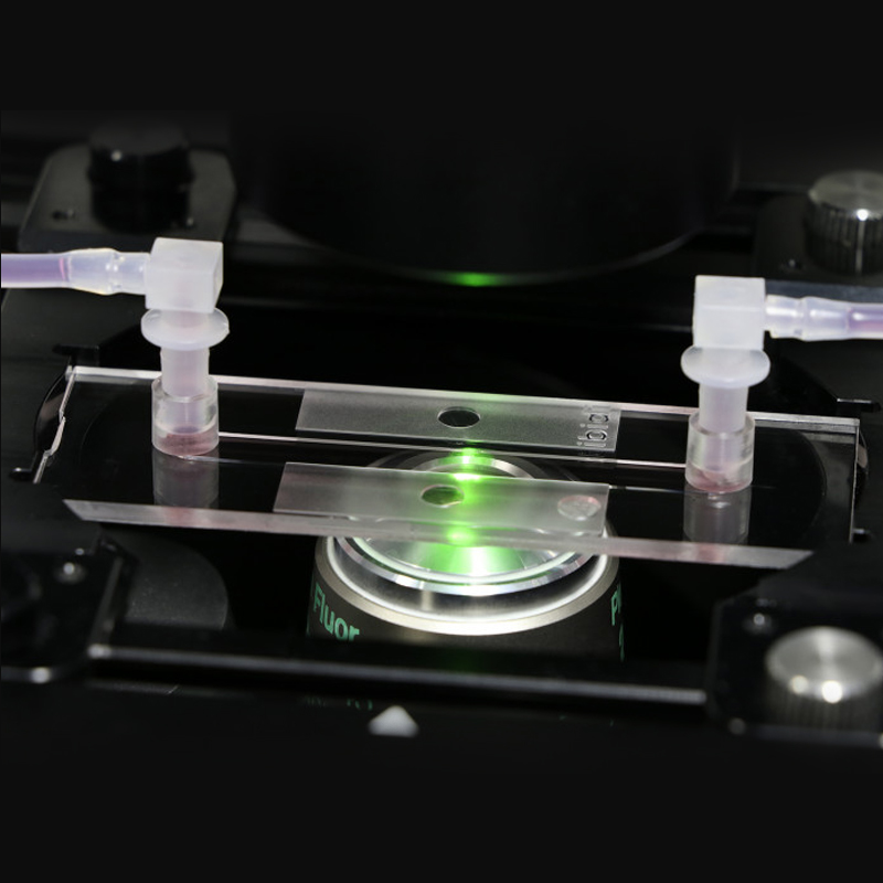
* Contains Luer adapters for easy tube and pump connection (e.g., to the ibidi Pump System)
* ibidi Polymer Coverslip on top and bottom for high-resolution light microscopy
* Partially compatible with upright microscopy
* Defined shear stress levels
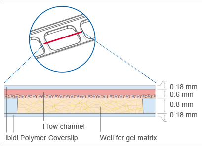
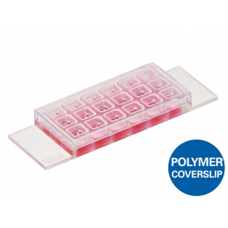
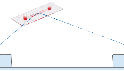
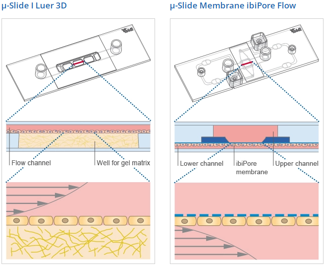
The Principle of the μ-Slide I Luer 3D
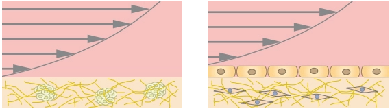
The μ-Slide I Luer 3D has three wells with one channel on top for culturing cells on a 3D gel matrix with defined flow. Each well can be filled with a gel, on which cells can be cultivated and microscopically investigated. The channel can be connected to a pump (e.g., to the ibidi Pump System) for the application of defined shear stress.
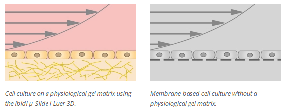
Using this method, an in vivo-like functional monolayer (e.g., an endothelial barrier) can be established on the gel matrix without any artificial filters or membranes involved. The μ-Slide I Luer 3D is especially designed for the simulation of blood vessels using endothelial cells.
Standard Filling Procedure
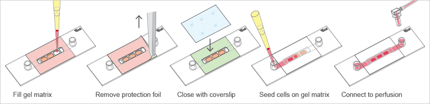
The μ-Slide I Luer 3D is a versatile channel slide for a huge variety of cell culture applications using a 3D gel matrix and shear stress:
Establishing a cell monolayer on the gel matrix—investigate potential polarization effects
Culturing cells or cell clusters inside the gel matrix
Performing assays using different gel matrices with varying concentration, stiffness, or chemical compounds
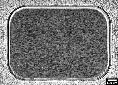
Stitched image of HUVECs in the 3 wells of the μ-Slide I Luer 3D on a Collagen Type I gel. The cells were cultured under a defined shear stress of 10 dyn/cm2 for 5 days. Phase contrast microscopy, 4x objective.
The μ-Slide I Luer 3D is suitable for a wide range of cell culture applications. The three wells can be filled with different gel matrices or different matrix concentrations. Cells can be cultivated as a monolayer on top of the gel. Alternatively, single cells or cell clusters can be embedded into the gel. The combination with the flow channel enables defined shear stress on the monolayer on the gel matrix. If cells are embedded inside the gel matrix, the perfusion delivers oxygen and nutrients to them.
These applications have been tested by the ibidi R&D team or by our customers.
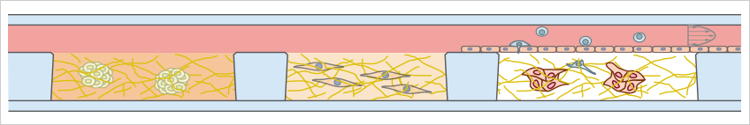
Endothelial Barriers Without Artificial Membrane
The μ-Slide I Luer 3D allows for creating an endothelial barrier without the need of an artificial filter membrane. Endothelial cells can be seeded on a suitable gel matrix, such as Collagen Type I, Rat Tail. After connecting the slide to a pump and applying defined shear stress, an in vivo-like endothelial barrier assay is created. This model simulates blood vessels on a soft ECM matrix instead of using a hard filter membrane.
Read our Application Note 60 for a specific protocol with HUVEC, Collagen Type I and the ibidi Pump System.
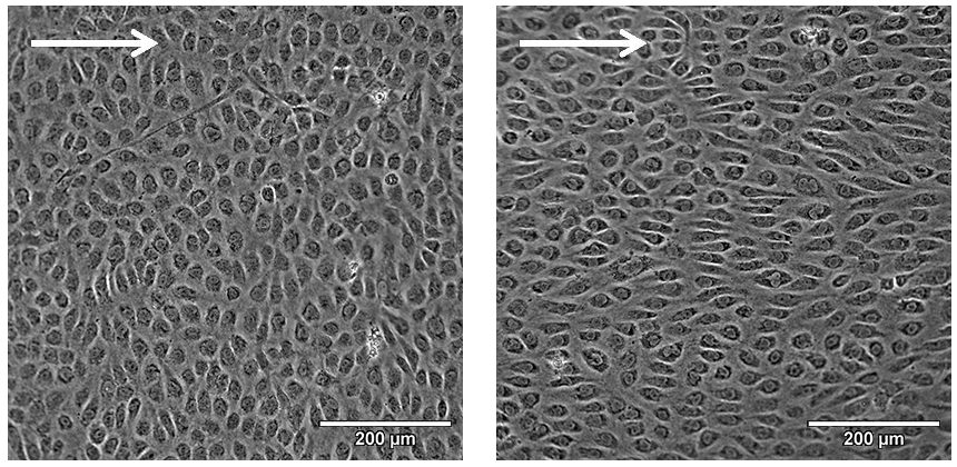
Phase contrast microscopy of HUVEC after culturing them under flow at 10 dyn/cm2 for 2 days (left) and 5 days (right) on a Collagen Type I rat tail (2 mg/ml). Note the cobblestone-like cell morphology after 5 days of culture under flow. 20x objective.
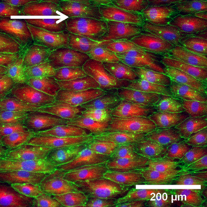
Fluorescence microscopy of HUVEC after culturing them under flow at 10 dyn/cm2 for 5 days on a Collagen Type I rat tail (2 mg/ml). Immunostaining of alpha-tubulin (red); the F-actin cytoskeleton was stained using phalloidin (green). Nuclei are stained with DAPI (blue). 10x objective.
Cell Polarization
A cell monolayer is seeded on a hydrogel and defined shear stress can be applied. Being compatible with all pipettable gel matrices, the μ-Slide I Luer 3D allows a wide variety of polarization studies (e.g., with epithelial cells).
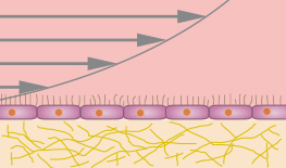
Polarized cells on a gel matrix, cultured under defined shear stress.
Potential Use
The following examples illustrate further potential product uses. ibidi has not yet tested these applications in-house, therefore we cannot provide specific protocols. However, from a technical point of view, these applications should be possible.
3D Cell and Tissue Assays
Several three-dimensional cell culture experiments can be performed using the μ-Slide I Luer 3D. For example, single cells or cell clusters can be seeded in the hydrogel matrix. Optionally, defined shear stress can be applied to a cell monolayer on top.
Spheroids (left) or single cells (right) embedded into a 3D gel matrix.
Rolling, Adhesion, and Transmigration Assays
For physiological rolling, adhesion, and transmigration approaches, the endothelial cell layer can be supplied with suspension cells on the luminal side and target cancer cells on the basal side. Optionally, defined shear stress can be applied.
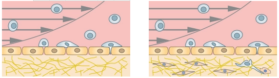
Co-culture of an endothelial cell layer and suspension cells for rolling and adhesion assays (left),
optionally with additional target cancer cells inside the extracellular matrix (right).
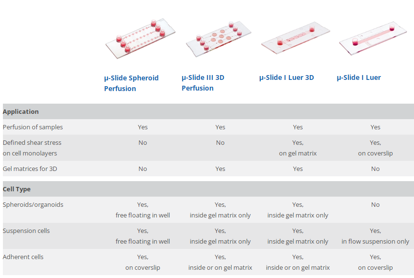
Decison Guide: ibidi Solutions for Two-Compartment and Membrane Assays
Slide principle
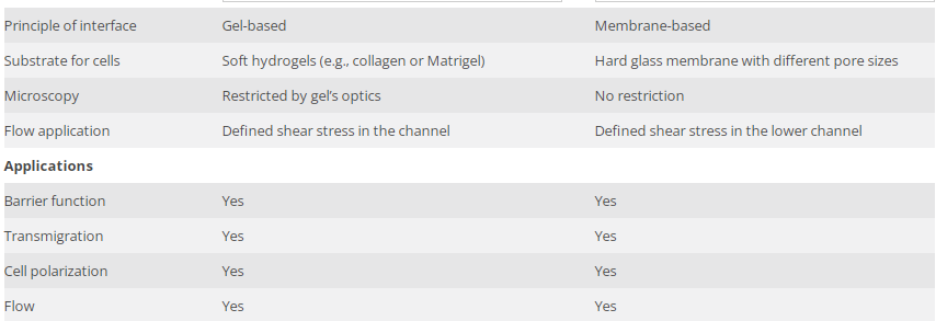
Which Slide Should I Use for My Application?